
The Kavli Institute for Nanoscience Discovery (KINsD) is a groundbreaking interdisciplinary science institute focused on world-class nanoscience research. Together, we are shaping the future of nanoscience, fostering an inclusive and collaborative KINsD working ethos, and driving breakthrough discoveries that have the potential to revolutionize our understanding of the world around us.

Our Research Themes
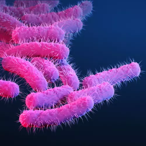
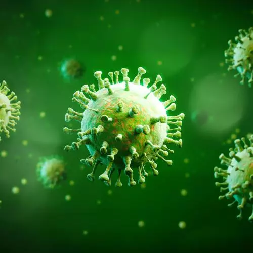
This is a medical illustration of drug-resistant, Shigella sp. bacteria

Antimicrobial Resistance

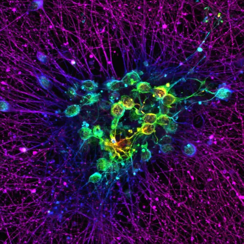
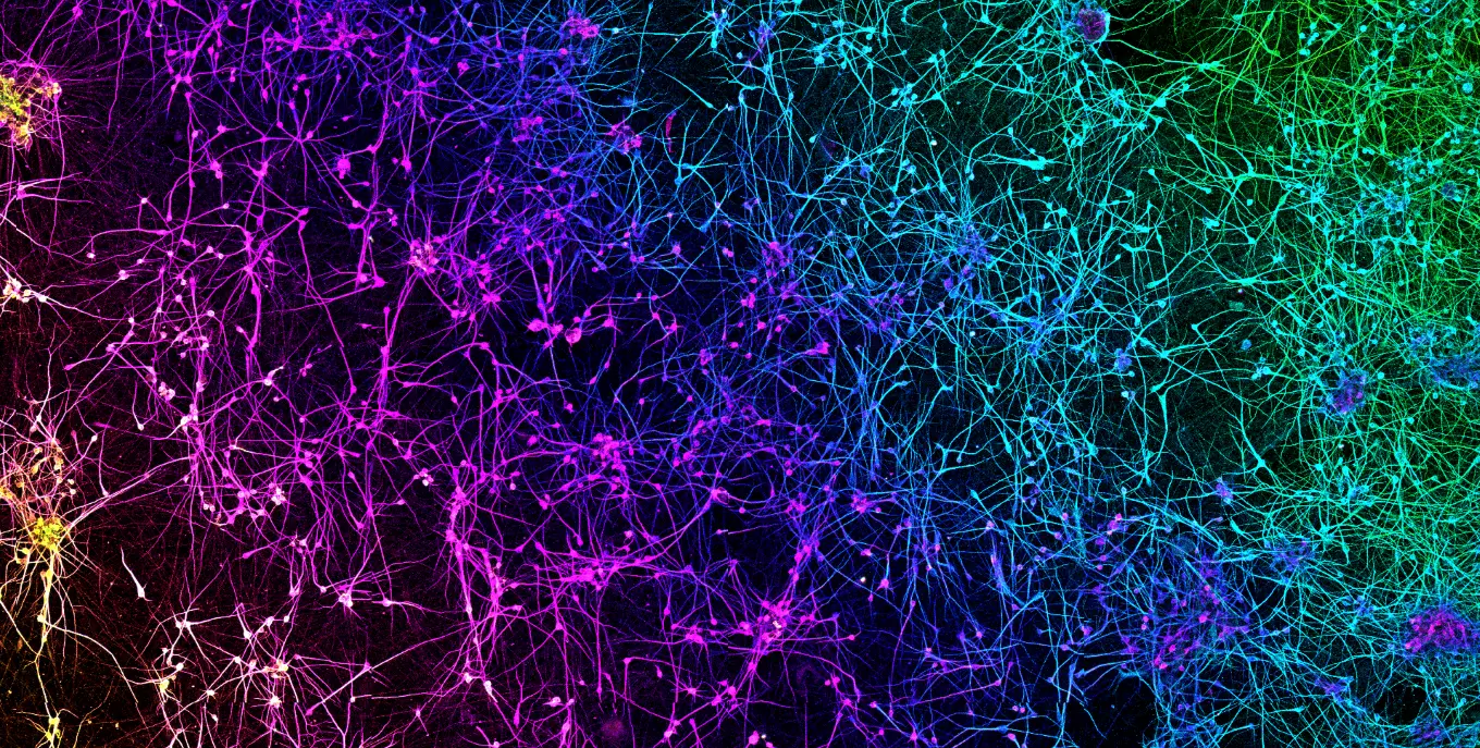
Fluorescence microscopy image of a dense neural network, with branching fibres glowing magenta and purple around clustered cells highlighted in green, yellow and cyan against a black background.

Brain Health

infection disease

Infectious Diseases

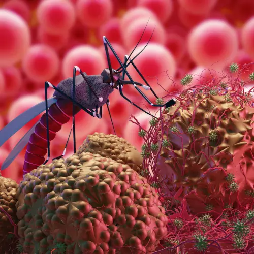
an image of a mosquito standing on blood molecules.

Malaria

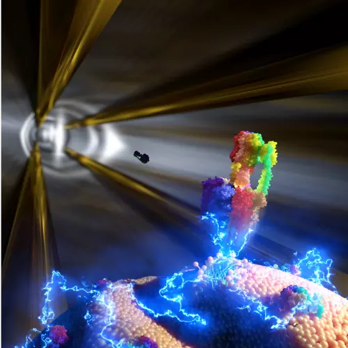
Scientific illustration of a colourful protein molecule interacting with a cell membrane, surrounded by electric-blue energy effects and dramatic golden light beams against a dark background.

New Tools for Nanoscience Discovery

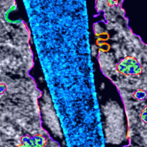
Microscopy illustration of a blue nanoneedle penetrating a cell, with surrounding cellular structures highlighted in grey, green, pink, orange and purple.

Next Generation Biomaterials

Our Research Facilities and Expertise

Shared research facilities are at the heart of the KINsD. We house state-of-the-art facilities for cryo-EM, super-resolution microscopy, mass photometry and mass spectrometry. 

In addition to being open for use by all of our Kavli Oxford members, the facilities can be accessed by researchers from outside our institute or outside the University of Oxford.
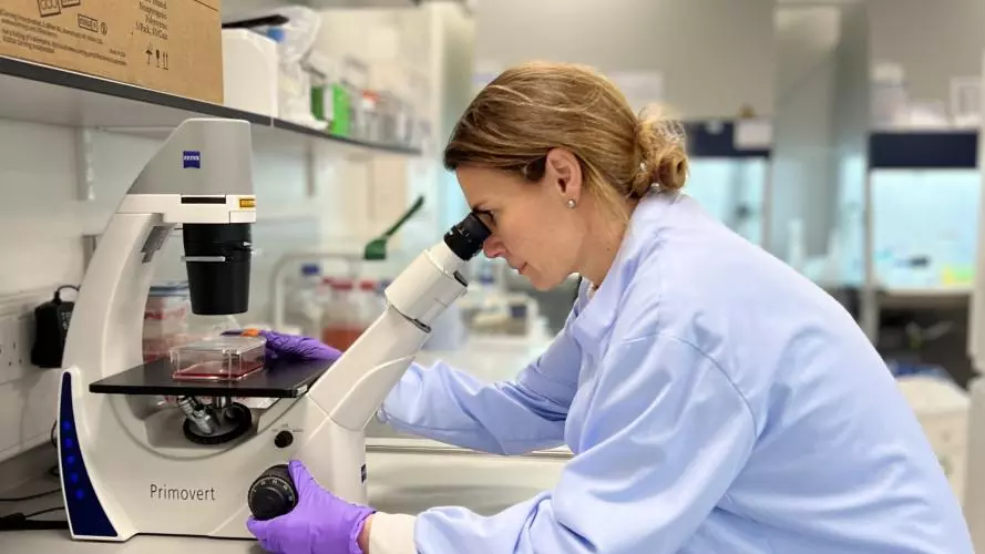
Professor Esther Becker working in the lab.
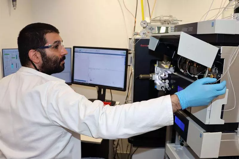
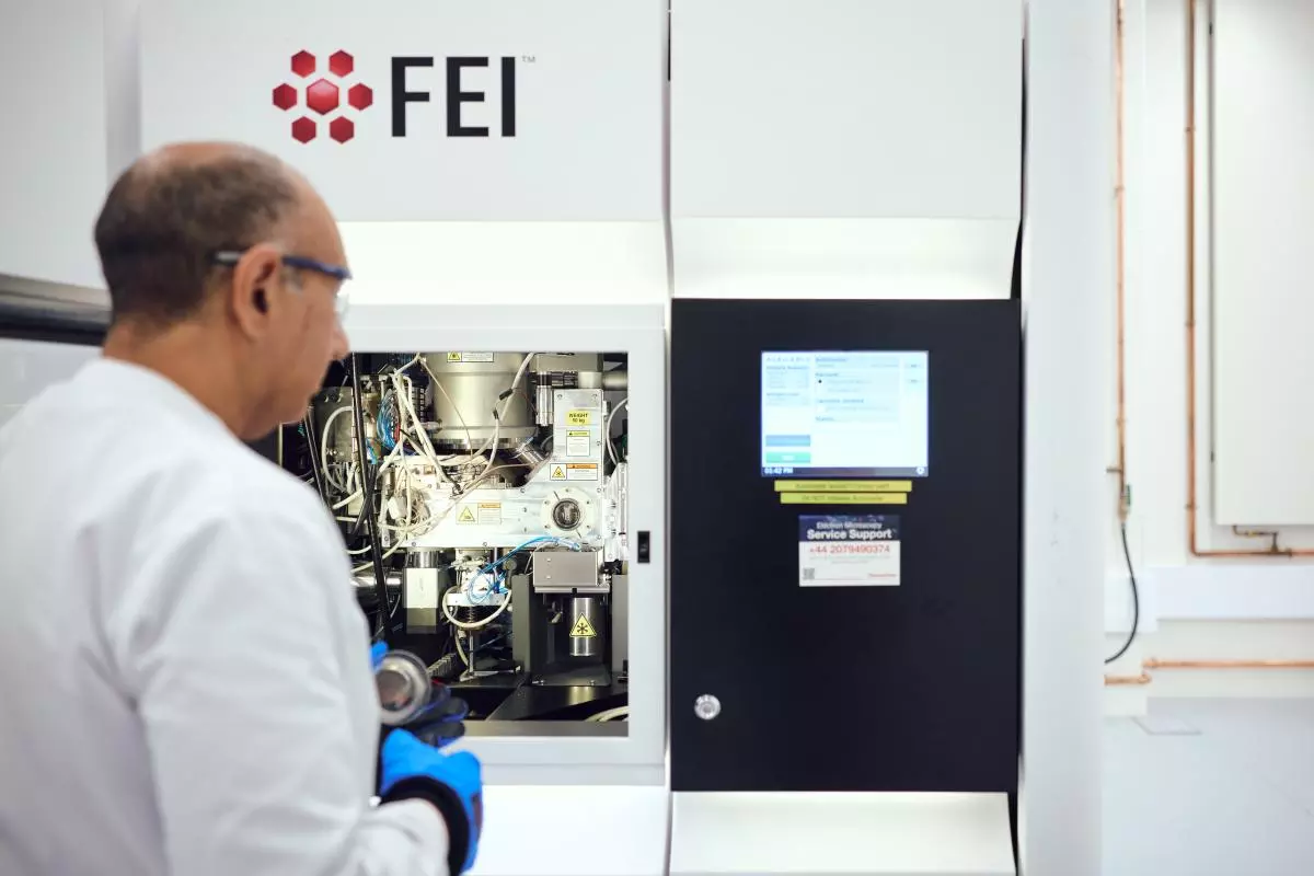
A scientist wearing blue gloves operates a large cryo-EM in a laboratory, inspecting its internal components beside a digital control screen.

Cryo-EM facility

Oxford Nanoimager (ONI)
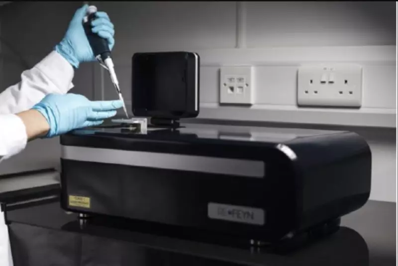
Oxford Nanoimager (ONI)

Mass Photometry

Mass photometry

Mass Spectrometers

Mass spectrometers

Latest News

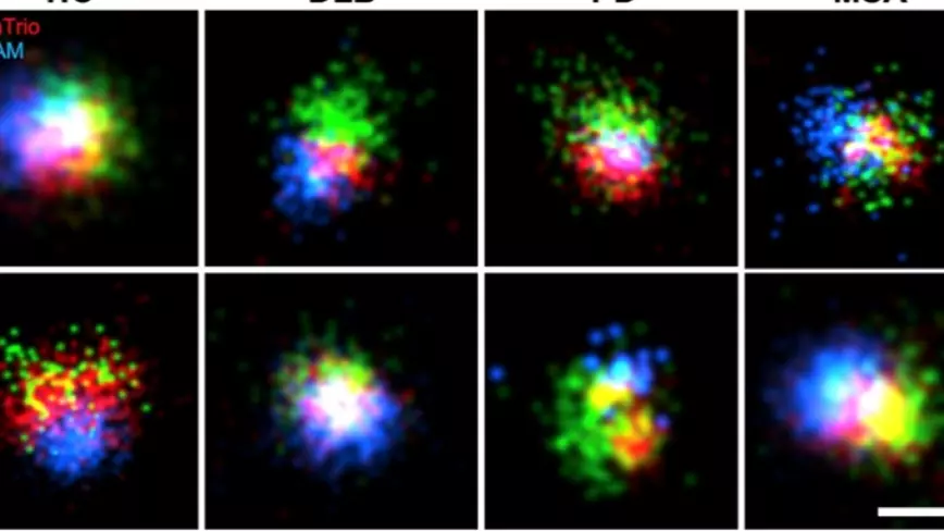
Fluorescence microscopy images comparing C-terminal and N-terminal alpha-synuclein staining across healthy controls (HC) and samples from DLB, Parkinson’s disease (PD), & multiple system atrophy (MSA). Red, green, & blue signals show protein localisation

Kavli Oxford researchers reveal differences in alpha-synuclein associated with neurodegenerative diseases

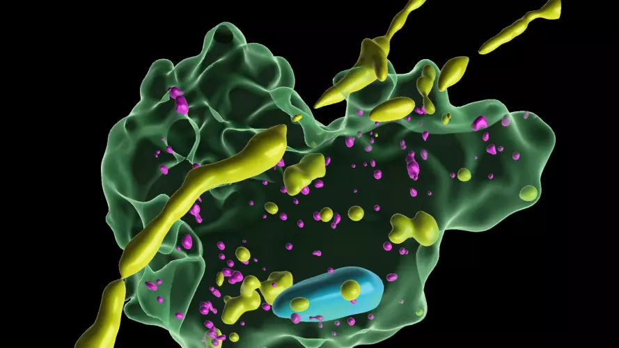
Scientific 3D illustration of a molecular structure featuring a translucent green protein surface, yellow and magenta molecules, and a blue component against a black background.

Kavli Oxford researchers discover how brain immune cells protect neurons by ‘nibbling’ away harmful material

Scientific illustration of a viral particle assembling from protein subunits on a dark blue background.

Kavli Oxford researchers capture the step-by-step assembly of individual virus-like particles

Upcoming Events

No items found.